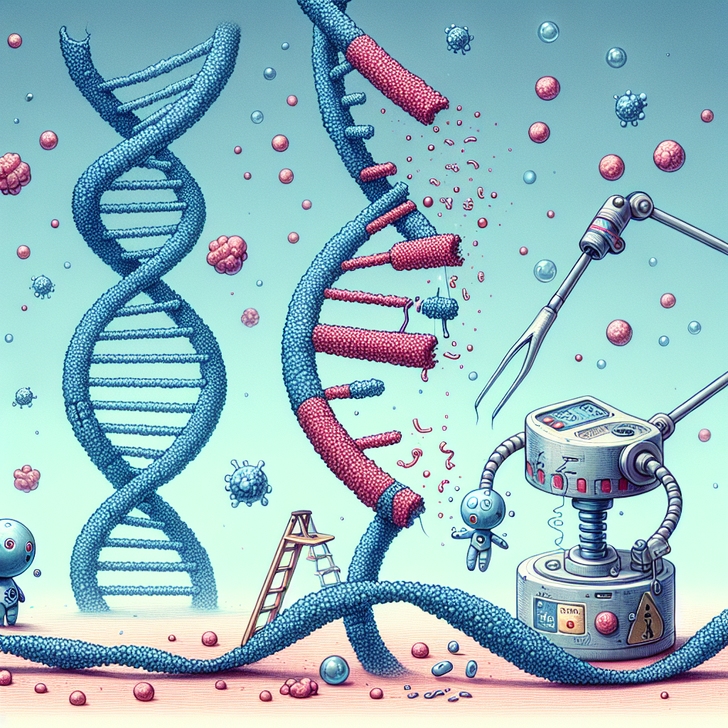
Understanding how a disease begins and identifying irregularities require examining various levels of biological organization, including DNA, chromosomes, cells, tissues, organs, and joints. Below is a step-by-step overview:

How the disease starts and identifying abnormalities:
Step 1: Genetic & Chromosomal Abnormalities
Chromosomes & DNA: Each human cell contains 23 pairs of chromosomes made up of tightly packed DNA. DNA carries genes, the instructions for producing proteins that regulate cell function.
• Mutations: Diseases often begin when a mutation occurs in DNA. This can be inherited (genetic diseases like cystic fibrosis) or acquired due to environmental factors (such as radiation or toxins). Mutations may involve:
• Point mutations (single nucleotide changes).
• Insertions or deletions (extra or missing DNA sequences).
• Chromosomal abnormalities (missing or extra chromosomes, like in Down syndrome—trisomy 21).
Step 2: Cellular Changes & Dysfunction
Gene Expression & Protein Production: Mutations can lead to improper protein function or production. This affects the cell’s ability to repair, divide, or communicate with other cells.
Abnormal Cell Behavior: Cells may:
- Grow uncontrollably (as in cancer).
- Become dysfunctional (as in neurodegenerative diseases like Parkinson’s).
- Fail to respond to signals (such as insulin resistance in diabetes).
Step 3: Tissue & Organ Impact
As defective cells accumulate, they form damaged or dysfunctional tissues.
For example:
- In autoimmune diseases, immune cells mistakenly attack normal tissues (e.g., rheumatoid arthritis in joints).
- In neurological diseases, faulty neurons lead to motor or cognitive dysfunction (e.g., Parkinson’s).
- In cardiovascular diseases, plaque buildup in blood vessels restricts oxygen to the heart.
Step 4: Organ Dysfunction & Symptoms
As tissues deteriorate, entire organs lose function:
- Liver disease may start with liver cell inflammation (hepatitis) and lead to fibrosis or cirrhosis.
- Kidney disease may begin with nephron damage and progress to kidney failure.
- Joint diseases like osteoarthritis start with cartilage breakdown, leading to inflammation, stiffness, and pain.
Step 5: Systemic & Whole-Body Effects
At advanced stages, diseases affect multiple organs and systems. Chronic inflammation, immune overactivation, and metabolic disruptions contribute to systemic conditions like diabetes, autoimmune disorders, and multi-organ failure.
DNA can be damaged by toxins or radiation through a series of molecular-level events that disrupt its structure and function. Here’s a step-by-step breakdown of how this happens:
Step 1: Exposure to Toxins or Radiation
• Toxins: These include heavy metals (mercury, lead, cadmium), pesticides, industrial chemicals, and cigarette smoke. They generate free radicals, reactive molecules that attack DNA.
• Radiation: Ultraviolet (UV) rays from the sun, X-rays, and gamma radiation can break DNA strands or cause mutations by energizing atoms in DNA molecules.
Step 2: Interaction with DNA
• Direct Damage: Radiation and some toxins can directly break the phosphate-sugar backbone of DNA, leading to single-strand or double-strand breaks.
• Indirect Damage: Free radicals generated by toxins or radiation steal electrons from DNA molecules, causing oxidative damage. This leads to abnormal chemical bonds, base modifications, and mutations.
Step 3: Types of DNA Mutations Caused
1. Point Mutations: A single base pair is substituted, inserted, or deleted. Example: A mutation in the p53 tumor suppressor gene can lead to uncontrolled cell growth (cancer).
2. Frameshift Mutations: Deletion or insertion of DNA bases shifts the reading frame, causing major changes in protein production.
3. Chromosomal Abnormalities: Toxins or radiation can cause large-scale damage, such as:
• Deletions: Loss of large DNA sections.
• Duplications: Extra copies of DNA segments.
• Translocations: DNA segments break off and attach to different chromosomes.
• Inversions: DNA segments flip within the same chromosome.
Step 4: Cellular Consequences
Failed Repair Mechanisms: Normally, cells repair damaged DNA using enzymes like DNA polymerase. However, severe damage overwhelms repair mechanisms, leading to:
• Senescence: The cell stops dividing permanently.
• Apoptosis: The cell self-destructs to prevent further harm.
• Uncontrolled Growth: If the damage affects tumor suppressor genes (e.g., p53) or oncogenes, the cell may grow uncontrollably, leading to cancer.
Step 5: Tissue and Organ Dysfunction
When enough cells are damaged or die, the affected tissue loses function.
Examples:
• Liver disease from toxin-induced DNA mutations leads to fibrosis and liver failure.
• Neurodegenerative diseases (e.g., Parkinson’s, Alzheimer’s) can arise from oxidative DNA damage in brain cells.
• Cancer develops when DNA damage leads to unregulated cell growth.
Repairing DNA damage in an orderly and efficient manner is critical for maintaining genomic stability and preventing diseases like cancer. Cells have evolved sophisticated repair mechanisms to address different types of DNA damage.
Below is a step-by-step explanation of how DNA repair occurs, integrating the quantum physics principles discussed earlier and emphasizing the orderly nature of these processes.
Step 1: Detection of DNA Damage
1. Sensors: – Specialized proteins (e.g., ATM, ATR, PARP) detect DNA damage by recognizing abnormal structures (e.g., breaks, crosslinks, or modified bases).
Quantum Sensing: These proteins may use quantum effects like electron tunneling to detect subtle changes in DNA structure.
2. Signal Amplification: – Damage detection triggers a cascade of signals to recruit repair machinery. —
Step 2: Types of DNA Repair Mechanisms
Cells use different repair pathways depending on the type of damage:
1. Base Excision Repair (BER) – Damage Type: Single-base modifications (e.g., oxidation, alkylation).
Steps:
- 1. Glycosylase Enzymes: Recognize and remove the damaged base, creating an abasic site.
- 2. AP Endonuclease: Cuts the DNA backbone at the abasic site.
- 3. DNA Polymerase: Fills the gap with the correct nucleotide.
- 4. DNA Ligase: Seals the backbone.
Quantum Aspect: Glycosylases use quantum tunneling to locate and excise damaged bases efficiently.
2. Nucleotide Excision Repair (NER)
Damage Type: Bulky lesions (e.g., thymine dimers, chemical adducts).
Steps:
- 1. Damage Recognition: Proteins like XPC and XPA identify the lesion.
- 2. Helicase Unwinding: TFIIH helicase unwinds the DNA around the damage.
- 3. Endonuclease Cleavage: XPF and XPG cut the damaged strand on either side of the lesion.
- 4. DNA Polymerase: Synthesizes a new strand using the undamaged strand as a template.
- 5. DNA Ligase: Seals the backbone. – Quantum Aspect: The unwinding and cutting processes involve energy transfer and conformational changes driven by quantum effects.
3. Mismatch Repair (MMR) Damage Type: Base-pair mismatches or insertion/deletion loops. –
Steps:
- 1. Recognition: Mismatch recognition proteins (e.g., MSH2, MSH6) bind to the error.
- 2. Excision: Exonucleases remove the mismatched segment.
- 3. Resynthesis: DNA polymerase fills the gap.
- 4. Ligation: DNA ligase seals the backbone.
Quantum Aspect: Mismatch recognition involves quantum-level interactions between proteins and DNA.
4. Double-Strand Break Repair (DSBR): Damage Type: Breaks in both DNA strands.
Pathways: Homologous Recombination (HR):
- 1. Resection: Exonucleases process the broken ends to create single-stranded overhangs.
- 2. Strand Invasion: RAD51 protein facilitates pairing with a homologous sequence (e.g., sister chromatid).
- 3. DNA Synthesis: The undamaged strand serves as a template for repair.
- 4. Resolution: The repaired strands are resolved and ligated.
Non-Homologous End Joining (NHEJ):
- 1. End Recognition: Ku70/Ku80 proteins bind to the broken ends.
- 2. Alignment: DNA-PKcs aligns the ends.
- 3. Ligation: XRCC4 and DNA ligase IV seal the break.
Quantum Aspect: Strand pairing in HR and end alignment in NHEJ involve quantum-level interactions.
Step 3: Coordination of Repair Pathways
- 1. Cell Cycle Checkpoints: – DNA damage activates checkpoints (e.g., G1/S, G2/M) to halt the cell cycle until repair is complete.
- 2. Protein Interactions: – Repair proteins form complexes (e.g., repairosomes) to coordinate their activities.
- 3. Quantum Coherence: – Energy transfer and electron tunneling may facilitate communication between repair proteins.
Step 4: Error Prevention and Quality Control
- 1. Proofreading: – DNA polymerases have proofreading activity to correct errors during repair.
- 2. Cross-Talk Between Pathways: – If one pathway fails, others can compensate (e.g., NHEJ as a backup for HR).
- 3. Apoptosis: – If damage is irreparable, cells undergo programmed cell death to prevent mutations.
Step 5: Applications in Medicine
- 1. Targeted Therapies: – Drugs like PARP inhibitors exploit DNA repair deficiencies in cancer cells.
- 2. Gene Editing: – CRISPR-Cas9 uses cellular repair mechanisms to introduce precise genetic changes.
- 3. Radiation Therapy: – Enhances DNA damage in cancer cells while sparing normal cells.
Quantum Physics in DNA Repair
- 1. Quantum Tunneling:– Enzymes like DNA photolyase use tunneling to transfer electrons and repair UV-induced damage.
- 2. Energy Transfer: – Repair proteins use resonance energy transfer to locate damage efficiently.
- 3. Conformational Changes: – Quantum-driven dynamics enable repair proteins to adopt the correct shapes for their functions.
Conclusion:
DNA repair is a highly orderly process that integrates molecular biology and quantum physics. Cells use specialized pathways to detect and repair damage, ensuring genomic stability. Understanding these mechanisms, including the quantum effects involved, is crucial for developing therapies to combat diseases caused by DNA damage.

Leave a Reply